In my opinion, the greatest concern in thyroid cytology is not
the distinction between follicular adenoma and carcinoma but the
distinction between a non-tumorous hyperplastic nodule and a follicular
tumor. Regarding the former problem, we have to accept that this
distinction cannot be made cytologically. Moreover, a follicular
adenoma is also a tumor and therefore it grows continously. Therefore
to send a patient with a benign tumor to surgery has only limited
disadvantage for the patient than to initiate surgery in a
non-neoplastic disease.
Regarding the latter, i.e. the distinction of a
non-tumorous lesion from a follicular tumor would be a classical task
of a
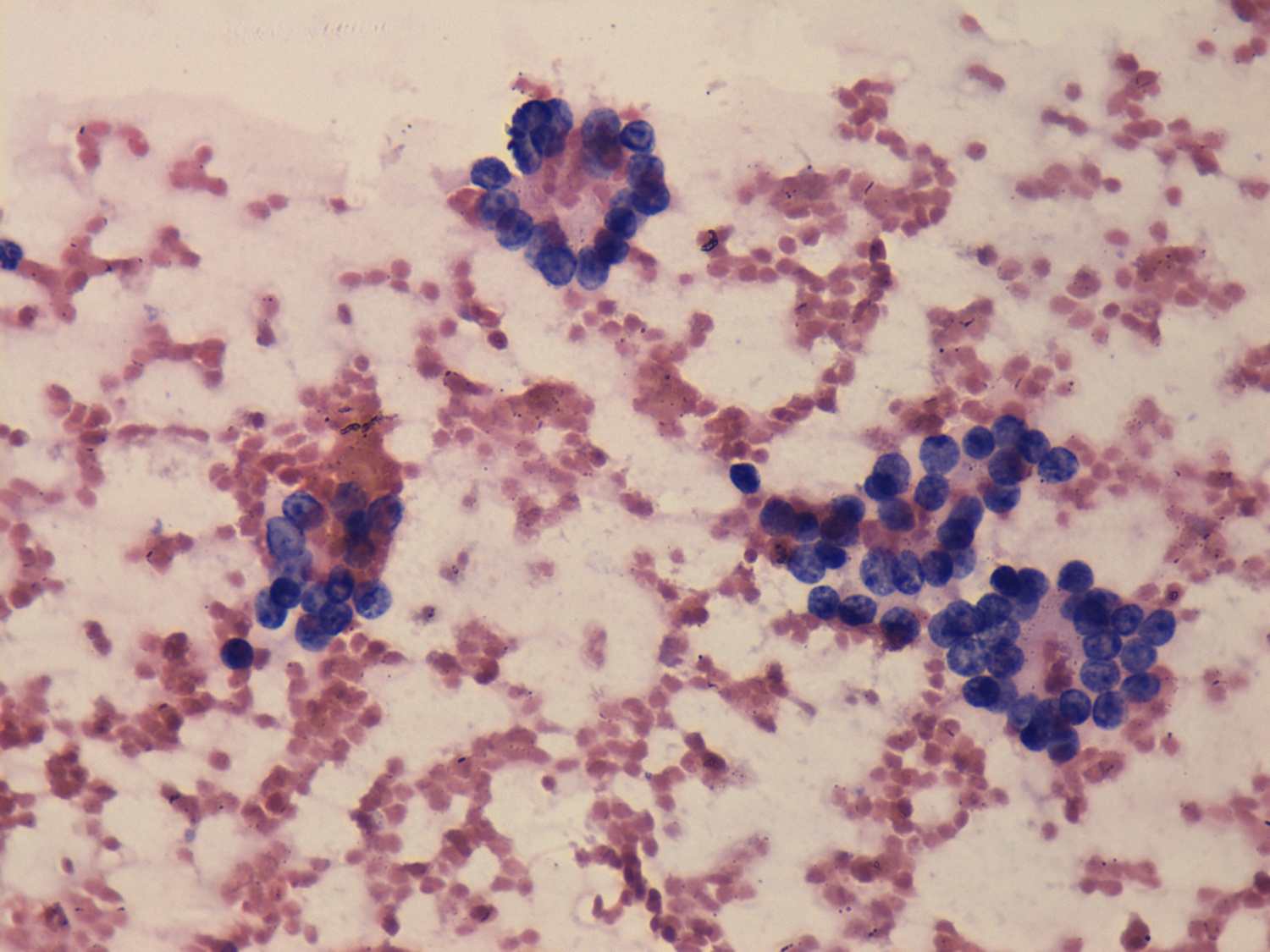
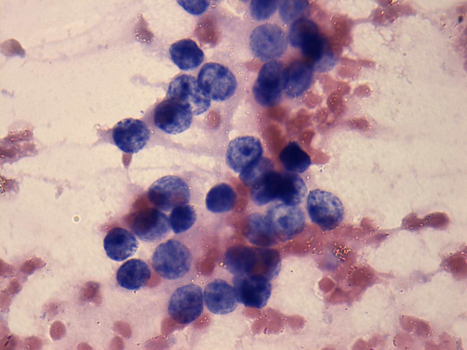
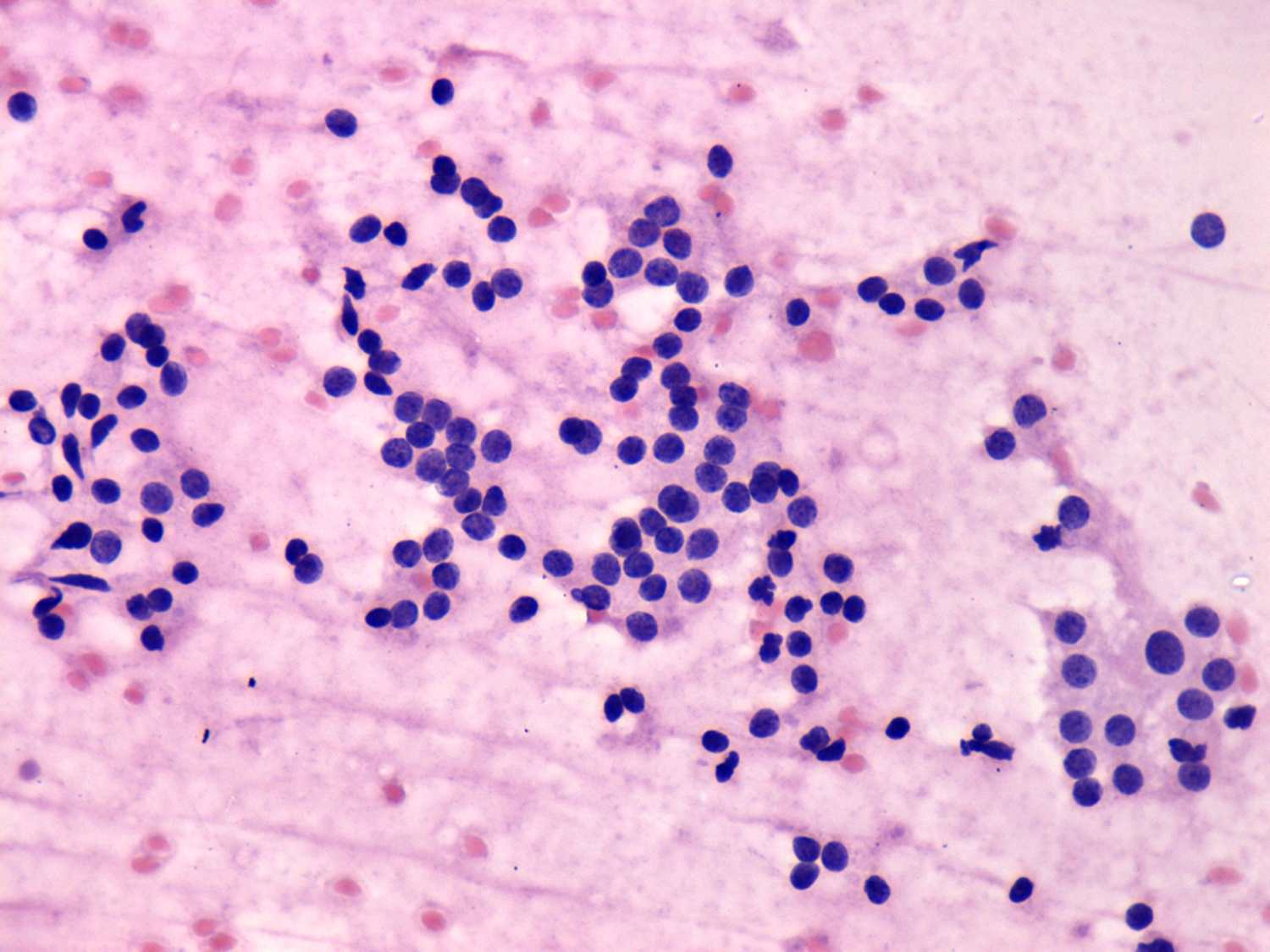
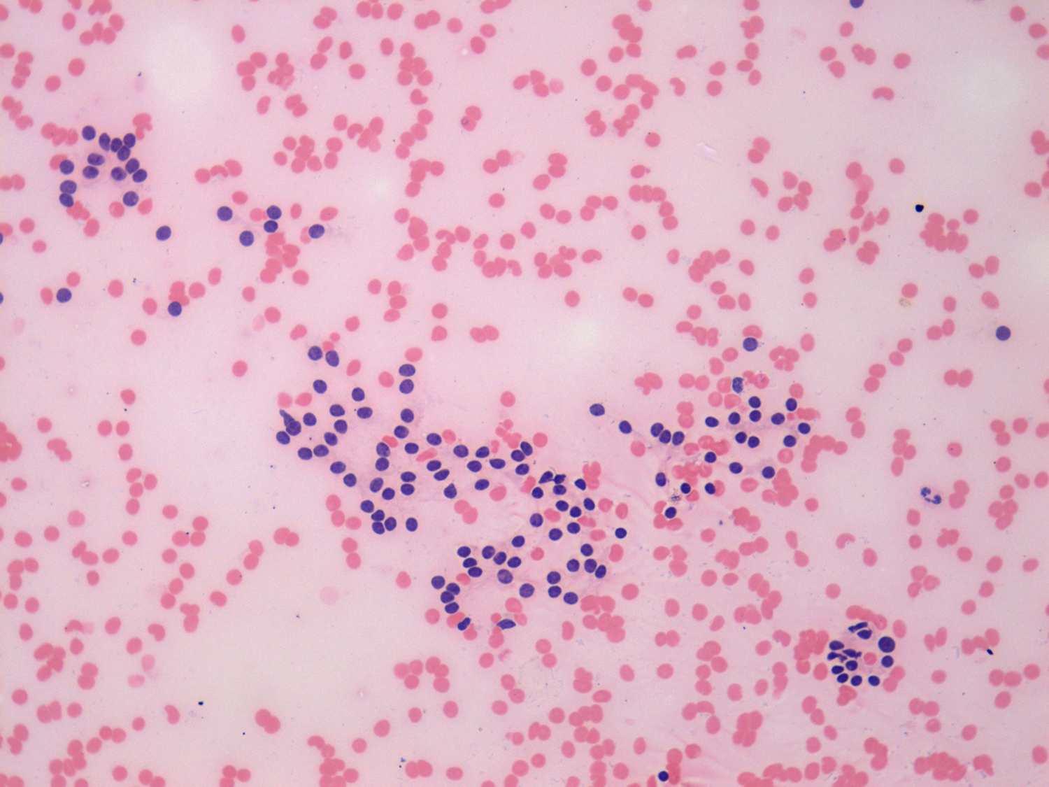
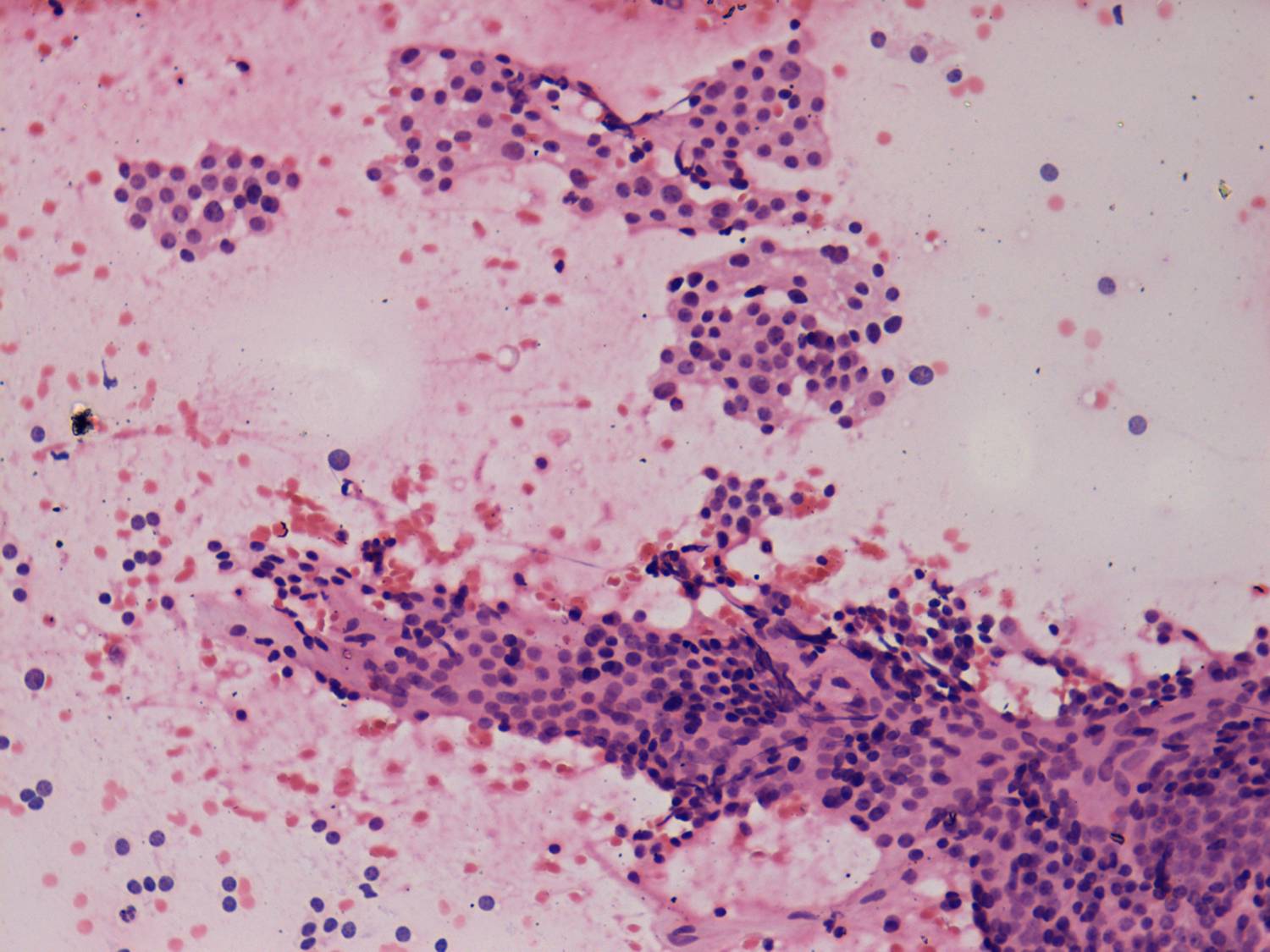
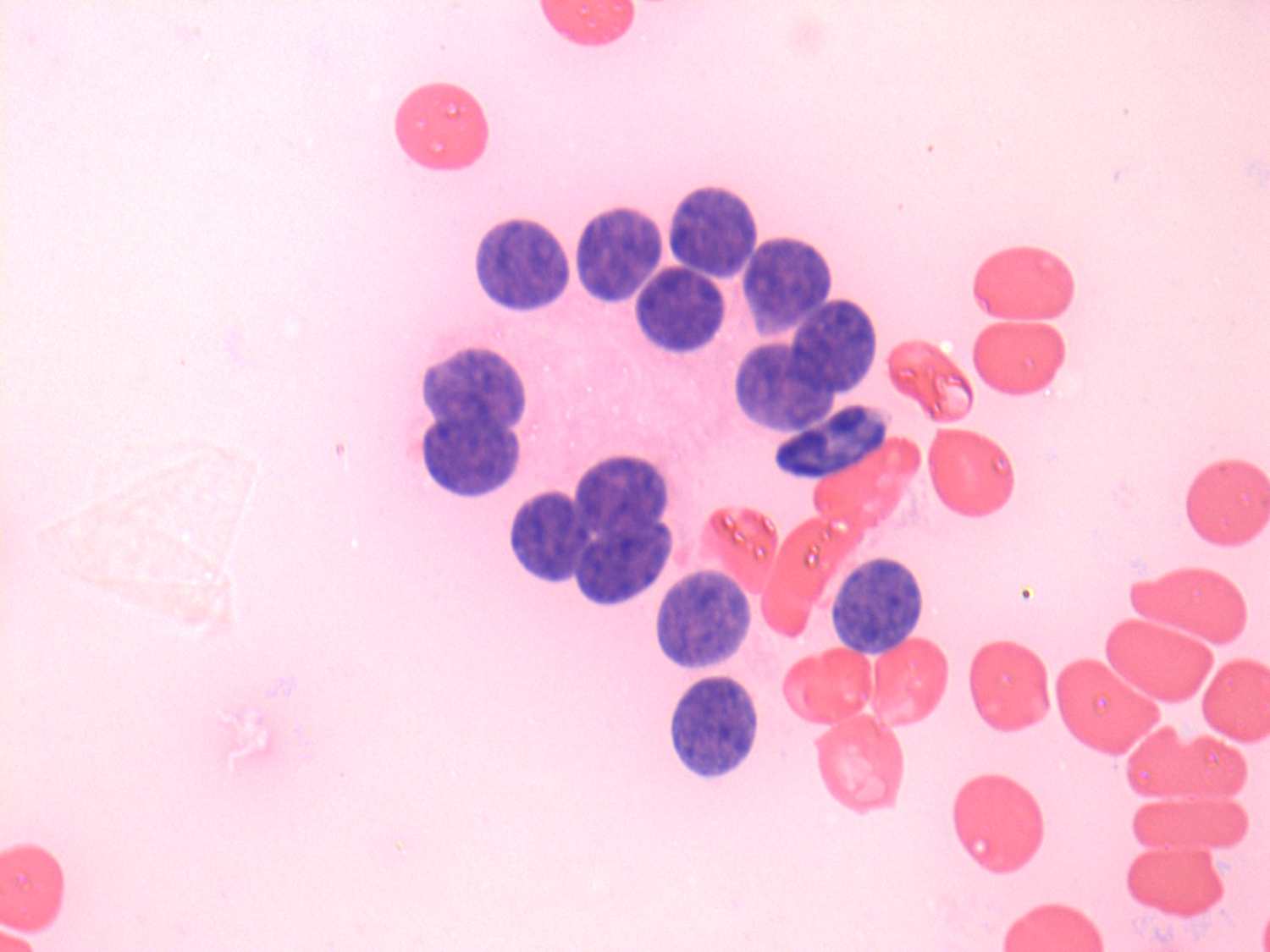
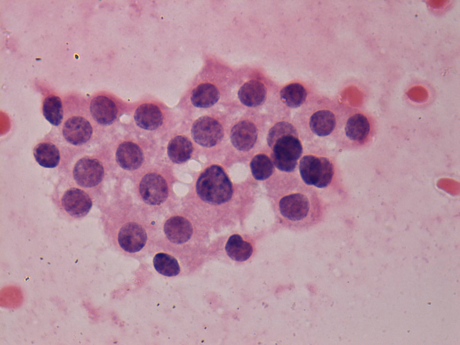
thyroid cytopathologist. First of all, the presence of colloid, the
ratio of microfollicles to normofollicles the ratio of follicles to monolayered sheets and the lack of
prominent nucleoli are of help. In fact, on thorough microscopic
analysis we can give an estimation about the risk being the lesion a
follicular tumor or not. In the everyday practice our estimation is in
the range of 10 to 80%. Not infrequently we are not able to fulfill
our obligation. The cases demonstrated above belong to this category.
Nevertheless, taking the sonographic
pattern into account is the most important clue not to operate
unnecessarily a patient with a non-tumorous lesion presenting
microfollicular proliferation. For details see later. In more than 95%
of follicular tumors,
we are able to recognize sonographic signs of a capsule (i.e. either halo sign or perinodular blood flow) which per
definitionem surrounds a follicular tumor. Benign hyperplastic nodules present less frequently halo sign or perinodular blood flow. Conversely, the lack of
sonographic signs of capsule significantly decreases the possibility of
a follicular tumor. It means that the risk of a follicular tumor is less than 5% in the event of microfollicular proliferation if the nodule lacks sonographic signs of a capsule.